
Last updated on September 4th, 2024 at 02:03 pm

Take Home Points
- For young adult patients with a high suspicion of an uncomplicated kidney stone (typical presentation) and adequate pain relief, CT can be avoided (even without prior stone history)
- CT can be avoided in middle aged patients with prior history of stone and typical presentation
- In older patients, CT should generally be obtained to rule out alternative diagnoses
- Pregnant and pediatric patients should have a radiologist performed/interpreted ultrasound
- POCUS can help in atypical or less typical presentations
Case
A 20 year old male with no past medical history comes back from the waiting room in excruciating pain in his right flank travelling into his groin that started abruptly while he was at work about an hour ago. He’s never had pain like this before, and he is nauseous from all the pain, and vomited once. He’s had no abdominal trauma, no surgeries. His vitals are all normal. He has no abdominal tenderness on exam, no CVA tenderness, and his exam is otherwise unremarkable.
This is an actual case I had earlier this year, and had a medical student see the patient (after I had started the workup and ordered pain control). When the medical student came back and presented the diagnosis we were both convinced of – ureterolithiasis – we came to the question “So do we need to image?”. My practice up until this point has been to get a CT scan without contrast in people who are having their first kidney stone, but starting my PGY3 year has gotten me contemplative about why I do the things I do – and whether those things are in the best interest of the patient.
A Multispecialty Panel Consensus
A recent RREBEL Core Cast with the legendary Swami covers this topic, and references a paper that I will be using to inform my imaging decisions in renal colic from now on – Imaging in Suspected Renal Colic: Systematic Review of the Literature and Multispecialty Consensus.1,2 Although the paper is a review, it’s the “multispecialty” part that stands out to me – a panel of physicians from the American College of Emergency Physicians, American College of Radiology, and the American Urological Association came together to discuss multiple common scenarios with variable consensus among the specialties. The panel was comprised of 9 providers, 3 EM physicians, 3 urologists, and 3 radiologists. So let’s break it down by clinical scenario…
But before that let’s define some things:
Age ranges: For the purposes of this article, I am defining pediatric as <18, young as 18-40, middle aged as 40-60, and elderly as 60+; the article does not define age ranges, rather each clinical scenario is repeated with a 35 year old (young), 55 year old (middle aged), and 75 year old (elderly) patient. These patients were all hypothetical, and although you can’t extrapolate the panel’s recommendations directly to patient care, there is a lot of value in seeing how both people within and outside of our specialty approach kidney stones.
Typical symptoms: Acute onset flank pain, nausea, vomiting, hematuria; Atypical symptoms: Subacute presentation (days), abdominal tenderness to palpation
History of kidney stone: History of prior diagnosis and spontaneous passage of a kidney stone
RDCT: Reduced-dose CT, basically, a CT that is protocolized to use less radiation when looking specifically for ureteral stones (this is not something we can order in our shop specifically, but may be something our radiologists are able to do behind the scenes – remember to include “rule out kidney stone” in your “Reason/Indication” box when ordering to give our radiologists some context); Non-con CT: CT abdomen & pelvis without contrast; POCUS: Point-of-care-ultrasound/Bedside ultrasound’ US: Ultrasound; formal US: An US performed by/interpreted by a radiologist
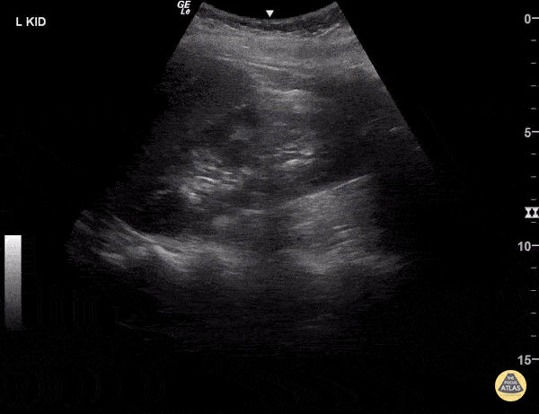
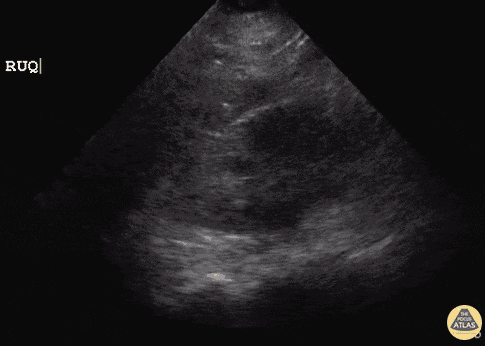
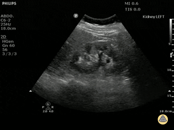
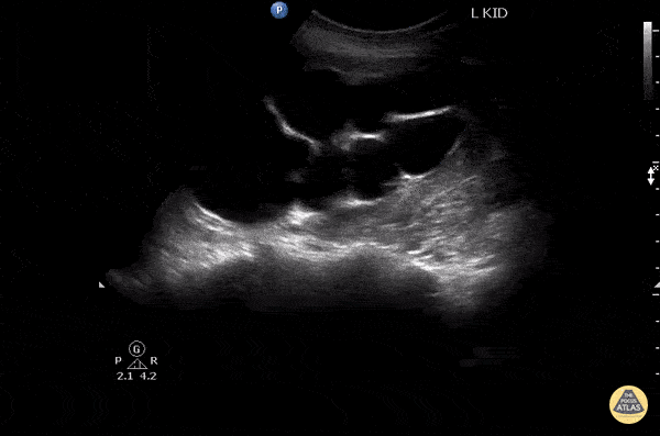
Wondering what hydronephrosis looks like on ultrasound? The POCUS Atlas has your back:3

Normal
Mild
Moderate
Severe
- Young Male Patients
- With a history of kidney stones
- Without a history of kidney stones
- Young Female Patients
- Middle Aged Male Patients
- Elderly Male Patients
- Pediatric Male Patient without a history of kidney stones, typical symptoms, pain controlled
Young male patient with history of kidney stones and typical symptoms, pain controlled
In this clinical scenario, the panel unanimously agreed that an initial CT was not needed. 4/9 actually recommended no imaging, while 5/9 recommended POCUS to look for hydronephrosis, though all agreed that regardless of your findings, you would not need further imaging. This begs the question of why you would need to do the scan in the first place, to which the panel responded that even if the POCUS did not influence a decision to obtain imaging, the ultrasound may provide a baseline in case they returned.
Bottom line: In a young male with prior stone history and typical presentation for a kidney stone with adequate pain control, consider POCUS as your first line imaging to look for hydronephrosis. Document your findings, but regardless of the findings, there is no need to obtain a CT scan.
Young male patient without history of kidney stones and with typical symptoms, pain controlled
The panel unanimously agreed on POCUS as initial imaging modality, with most of the panel without recommendation for further imaging regardless of the result. One respondent (one of the three EM physicians) recommended obtaining a non-con CT regardless of the POCUS findings essentially referencing the common teaching that every first time stone warrants a CT scan. One one hand, this sounds like dogmatic teaching rearing its ugly head, but on the other, this EM physician may be coming from a negative outcome or near miss where a CT scan would have made a difference. The decision ultimately comes down to how risk averse the individual provider is, but with eight other members of the panels (including 2 EM physicians, all 3 urologists, and 3 radiologists), I lean towards no further imaging.
Bottom line: In a young male with no prior stone history and typical presentation for a kidney stone with adequate pain control, consider POCUS as your first line imaging to look for hydronephrosis. Document your findings, but regardless of the findings, there is no need to obtain a CT scan.
Young male patient without history of kidney stones and atypical symptoms, pain controlled
Almost the entire panel recommended RDCT in this case, with only 1 recommending POCUS.
Bottom line: In a young male without prior stone history but typical presentation for a kidney stone with adequate pain control, consider RDCT/non-con CT POCUS as your first line imaging to look for ureterolithiasis and hydronephrosis.
Young male patient without history of kidney stones and with typical symptoms, pain not controlled
Unanimous agreement that this patient would need a RDCT.
Bottom line: In a young male with no prior stone history and typical presentation for a kidney stone but without adequate pain control, consider RDCT/non-con as your first line imaging to look for ureterolithiasis and hydronephrosis.
Young male patient with history of kidney stones and atypical symptoms, pain controlled
7/9 of the panel recommended POCUS much like the patient with typical symptoms, 2 recommended RDCT. If the POCUS is negative, 5/9 recommended no imaging, while 4/9 recommended a RDCT.
Bottom line: In a young male with prior stone history but atypical presentation for a kidney stone with adequate pain control, consider POCUS as your first line imaging to look for hydronephrosis. Document your findings, and if negative for hydronephrosis, consider either no imaging given adequate pain control, or RDCT/non-con CT to look for ureterolithiasis and hydronephrosis. Alternatively, if you would likely end up getting a RDCT/non-con CT in the setting of not seeing any hydronephrosis on POCUS, consider starting with a RDCT/non-con CT.
Young male patient with diagnosed ureteral stone on CT the day prior presenting again for similar typical symptoms
Unanimous – no imaging.
Bottom line: In a young male who was recently diagnosed with a CT confirmed ureteral stone, and is now presenting with the same typical symptoms that they initially presented with, consider no imaging for the patient.
Young male patient underwent a shock wave lithotripsy 2 days prior with typical symptoms, pain controlled
Tough situation, though most recommend formal US (6/9), 2 for RDCT, and 1 for POCUS.
Bottom line: In a young male who recently underwent shock wave lithotripsy, consider formal US or a RDCT/non-con CT.
Young male patient underwent stenting 2 days prior with typical symptoms, pain controlled
Unanimous agreement on POCUS.
Bottom line: In a young male who recently underwent ureteral stenting, consider POCUS.
Young female patient without history of kidney stones and with typical symptoms, pain controlled
8/9 agreed that the patient needs an US (6 for POCUS, 2 for formal US). One recommended RDCT.
Bottom line: In a young female with no prior stone history and typical presentation for a kidney stone with adequate pain control, consider POCUS or a formal US as your first line imaging to look for hydronephrosis.
Young pregnant female patient without history of kidney stones and with typical symptoms, pain controlled
Unanimous recommendation for US, with 8/9 for formal US over POCUS. Regardless of findings, the recommendation is for no further imaging.
Bottom line: In a young pregnant female with no prior stone history and typical presentation for a kidney stone with adequate pain control, consider formal US as your first line imaging to look for hydronephrosis.
Middle aged male patient with history of kidney stones and typical symptoms, pain controlled
5/9 on the panel recommended no imaging, 4/9 recommended initial POCUS.
Bottom line: In a middle aged male with prior stone history and typical presentation for a kidney stone with adequate pain control, consider POCUS as your first line imaging to look for hydronephrosis.
Middle aged male patient without history of kidney stones and with typical symptoms, pain controlled
Most (8/9) of the panel recommended initial RDCT.
Bottom line: In a middle aged male without prior stone history and typical presentation for a kidney stone with adequate pain control, consider RDCT/non-con CT scan of the abdomen and pelvis as your first line imaging to look for ureteral stone and hydronephrosis.
Middle aged male patient without history of kidney stones and with atypical symptoms, pain controlled
Unanimous agreement on getting a CT scan, but some variability of what type to obtain, 7 RDCT, 1 CT non-con, and 1 CT with IV contrast.
Bottom line: In a middle aged male without prior stone history and atypical presentation for a kidney stone with adequate pain control, consider RDCT/non-con CT scan of the abdomen and pelvis as your first line imaging to look for ureteral stone and hydronephrosis; if you have a high enough concern for alternative etiology, obtain a CT with IV contrast.
Elderly male patient with history of kidney stones and typical symptoms, pain controlled
Some variability here, though most recommended RDCT (7/9), 1 recommended POCUS, and 1 recommended a radiologist performed US.
Bottom line: In an elderly male with prior stone history and typical presentation for a kidney stone with adequate pain control, consider RDCT/non-con CT scan of the abdomen and pelvis as your first line imaging to look for ureteral stone and hydronephrosis.
Elderly aged male patient without history of kidney stones and with typical symptoms, pain controlled
Unanimous recommendation here for a non-con CT.
Bottom line: In an elderly male without prior stone history and typical presentation for a kidney stone with adequate pain control, consider RDCT/non-con CT scan of the abdomen and pelvis as your first line imaging to look for ureteral stone and hydronephrosis.
Elderly aged male patient without history of kidney stones and with atypical symptoms, pain controlled
Unanimous recommendation here CT with 5/9 recommending non-con CT and the rest recommending IV contrast.
Bottom line: In an elderly male without prior stone history and atypical presentation for a kidney stone with adequate pain control, consider a CT scan either with or without contrast to look for ureterolithiasis and hydronephrosis.
Pediatric male patient without history of kidney stones and with typical symptoms, pain controlled
Unanimous recommendation for ultrasound, 7/9 recommending formal US, 2 POCUS. No further imaging required if there is hydronephrosis, and if there isn’t, a formal US was recommended with 1/9 recommending a RDCT if there is no hydronephrosis on US.
Bottom line: In the pediatric male patient without history of prior kidney stones and typical symptoms with pain controlled, consider formal US as your first line imaging to look for hydronephrosis. Document your findings, regardless, there is likely no need for further imaging, but if there is no hydronephrosis, consider RDCT/non-con CT.
Author

Cite this post: Arman Hussain, MD. “Picture This: Imaging in Renal Colic”. GW EM Blog. August 9, 2023. Available at: https://www.gwuemblog.com/kidney-stone-imaging.
Related Posts:
rMETRIQ Score: Not yet rated/21
References
- 1.Swaminathan A. REBEL Core Cast 101.0 – Imaging in Renal Colic. REBEL EM. Published May 10, 2023. Accessed July 16, 2023. https://rebelem.com/rebel-core-cast-101-0-imaging-in-renal-colic/
- 2.Moore CL, Carpenter CR, Heilbrun ML, et al. Imaging in Suspected Renal Colic: Systematic Review of the Literature and Multispecialty Consensus. Journal of Urology. Published online September 2019:475-483. doi:10.1097/ju.0000000000000342
- 3.Hydronephrosis – TPA. The POCUS Atlas. Accessed July 21, 2023. https://www.thepocusatlas.com/hydro-and-obstruction